
3D Microscope:
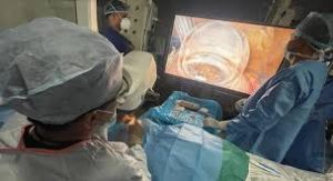
For the first time, the Indian Army’s Department of Ophthalmology at Army Hospital (Research and Referral), New Delhi, has successfully performed Minimally Invasive Glaucoma Surgery (MIGS) using a 3D Microscope.
- A microscope is an instrument that magnifies small objects, making them visible to the naked eye by bending (refracting) light rays through curved lenses.
- The most commonly used microscopes are optical microscopes, where visible light is focused through lenses to create an enlarged image.
- A 3D microscope produces images with depth information (X, Y, and Z axes), allowing researchers to visualize and measure the topography, volume, and internal structures of samples.
- Unlike traditional light microscopes, which provide flat, 2D images, 3D microscopes use advanced optical, electron, or computational techniques to capture and reconstruct three-dimensional data.
- This is particularly useful for studying complex biological or environmental samples, such as soil microbes, aquatic organisms.
- The 3D Microscope uses advanced three-dimensional visualisation, assisting in complex eye surgeries such as treatment for squint, cataract, corneal diseases, glaucoma, and retinal conditions.
- It employs special 3D polarisation glasses for surgeons and a 55-inch 4K ultra-HD display.
- Key advantages include:
- Reduced surgical time and lower complication rates compared to conventional microscopes.
- Decreased endoilluminator power requirements, thereby reducing photo-toxicity risks.
- Ease of performing surgeries in complex and rare cases.